Share |

-

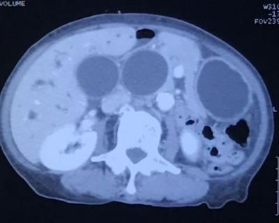
Biliary & Gastric Dilation
Figure 1: Computed tomography showing 3 homogenously enhanced lesions on axial view
-
Left Facial Tumour. What Is It?
Figure 1: Left facial tumour protruding from the orbital cavity displacing the eyeball downwards (eyeball not seen in picture)
On the Occasion of 10th Anniversary
Statin: Hope for a New Agent to Treat Osteoporosis
Vein of Galen Malformation
Figure 1: MRI of the brain in sagital T2WI and contrast-enhanced T1 MPRAGE demonstrate multiple serpiginous flow voids (star and bent arrow) representing dilated venous channels. Abnormal vessels (bent arrow) are seen draining into a dilated vein (thin arrow), which in turn drains into the superior sagital sinus. Narrowing of aqueduct of sylvius is marked with an arrowhead.
Pharmacovigilance and Reporting Adverse Drug Reactions
The History of RNAi and MicroRNA Discovery
Is Bariatic Surgery The Cure for Diabetes Mellitus?