Hello Visitor!
Log In
or
Register
Home
My profile
Home
About
About M&H
Aims and Scope
Editorial Board
Reviewers
Issues
Current Issue
Browse Archive
Search
Article In Press
For Authors
Instruction To Authors
Online Submission
For Reviewers
Instruction To Reviewers
Contact
Reviewer Agreement Form
Search issue
MEDICINE & HEALTH
About
Aims and Scope
Editorial Policies and Guidelines
Editorial Board
Issues
Current Issue
Browse Archive
For Authors
Guidelines for Authors
Submit manuscript
Keyword guideline (MeSH)
Guidelines for online submission
For Reviewers
Review Comments Form
Reviewer Agreement Form
Guide for Reviewer
Related Links
Universiti Kebangsaan Malaysia
UKM Medical Centre
Centre for Research and Instrumentation Management
Centre for Graduate Management
Home
Share
|
-
-
Article |
December 31, 2019 - 10:23am
| By
HAYATI F
,
AZIZAN N
,
CHONG TH
,
NIK AMIN S
,
ZAINAL ABIDIN ZA
Related Terms:
-
Quiz
-
Article |
September 6, 2019 - 11:10pm
| By
-
Related Terms:
-
Conference
-
Article |
August 22, 2019 - 8:27am
| By
-
Related Terms:
-
Conference
-
Article |
August 14, 2019 - 11:37pm
| By
HAYATI F
,
AZIZAN N
,
KADIR F
,
ANDEE DZ
,
SIBIN R
Related Terms:
-
Quiz
-
Article |
August 13, 2019 - 3:52pm
| By
IMA NIRWANA S
,
Srijit D
Related Terms:
-
Editorial
-
Article |
August 8, 2019 - 10:20pm
| By
MAHDY ZA
,
Srijit D
Related Terms:
-
Editorial
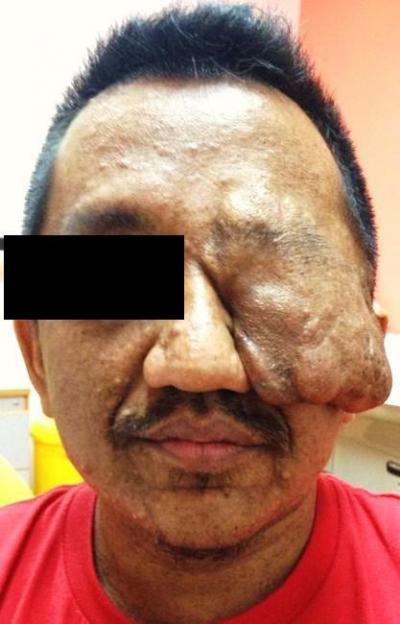
Left Facial Tumour. What Is It?
Article |
July 30, 2019 - 11:50pm
| By
GENDEH HS
,
Imran FH
,
Kosai NR
,
Gendeh BS
,
Ramzisham AR
,
Kelly EG
Related Terms:
-
Quiz
Innovations in Obesity and Metabolic Surgery: Boon or Bane?
Article |
July 25, 2019 - 10:35am
| By
REYNU R
,
KOSAI NR
Related Terms:
-
Editorial
splenic hilar mass with carcinoid symptom
Article |
June 28, 2019 - 12:29pm
| By
HAMIZI MH
,
SANDRASECRA S
,
HAYATI F
,
AZIZAN N
,
MAHAMAD DOM SA
,
CHUAH JA
Related Terms:
-
Quiz
-
abdominal mass with haematuria
Article |
June 28, 2019 - 12:26pm
| By
HAYATI F
,
AZIZAN N
,
CHONG TH
,
NIK AMIN S
,
ZAINAL ABIDIN ZA
Related Terms:
-
Quiz
-
« first
‹ previous
1
2
3
4
5
6
7
next ›
last »